

In this series, we are highlighting some of our founder, Dr. Yelena Sheptovitsky’s, favorite manuscripts, publications, and news pieces all relating to the world of protein expression, life science, and CRO’s.
Today we look at the author manuscript from “Transfection by Electroporation” by Huntington Potter, Ph.D., Richard Heller, Ph.D., Published in final edited form Curr Protoc Mol Biol. 2003 May ; CHAPTER: Unit–9.3. doi:10.1002/0471142727.mb0903s62.

ABSTRACT
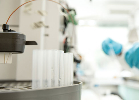
Electroporation–the use of high-voltage electric shocks to introduce DNA into cells–can be used with most cell types, yields a high frequency of both stable transformation and transient gene expression and, because it requires fewer steps, can be easier than alternate techniques. This unit describes electroporation of mammalian cells, including ES cells for the preparation of knockout, knockin, and transgenic mice,, , describes protocols for using electroporation in vivo to perform gene therapy for cancer therapy and DNA vaccination, and.outlines modifications for preparation and transfection of plant protoplasts.
One area that stands to be mentioned:
IN VIVO PROTOCOL
ELECTROPORATION INTO MUSCLE OR SKIN
Electroporation has been used successfully to deliver plasmid DNA to a variety of tissues in vivo (Heller et al., 2006a). Because of its physical nature, EP can be applied to practically any cell or tissue. Plasmid DNA in the appropriate diluent is injected into the tissue. Electrodes are then placed around the injection site and the cells within the tissue are subjected to a high-voltage electrical pulse of defined magnitude and length. The animals are then allowed to recover and the tissue is evaluated at specified time points following delivery. Factors that can be varied to optimize electroporation effectiveness are pulse width, number, amplitude and electrode configuration.
Materials—Animals to undergo procedure Syringe (1 CC) and needle size – 25–30 gauge
Linear or supercoiled, purified DNA preparation (see step 1)
Electrodes for administering the pulses
Electroporation power source
Additional reagents and equipment for harvesting tissue or evaluating expression levels and efficiency.
Anticipated Results
The efficiency of transfection by electroporation is dependent upon cell type. For fibroblasts, which are easily transfected by calcium phosphate or DEAE-dextran coprecipitation (UNITS 9.1 & 9.2), electroporation gives a stable transformation frequency of 1 in ~103 to 104 live cells—approximately that obtainable by the above traditional procedures. For cells refractory to traditional methods, electroporation gives a stable transformation frequency between 1 in 104 to 105 for most cell types. Occasionally a cell line (e.g., some T lymphocytes) will transfect poorly under our standard conditions (1 in 106), and even this frequency is sufficient to obtain significant numbers of transfectants. In general, cells that transfect efficiently for stable transformants also do so for transient gene expression. Increasing the number of cells and the amount of DNA used in the electroporation for studying transient gene expression can circumvent problems of low transfection efficiency and low promoter/enhancer efficiency.
For plant protoplast electroporation, the frequency of stable transformants is between 1 in 102 and 1 in 103 dividing cells.
Efficiency of in vivo electroporation is dependent on tissue type, protein being expressed, plasmid size and promoter. Skin delivery efficiency has been reported as high as 32%
(Heller et al., 2006b). Muscle has also been demonstrated to achieve efficient delivery of greater than 30% (Mir, et al, 1999).
To read the full manuscript, click here.
ARVYS Proteins, Inc. is a leader in the space of Antibody Development & Production. From generation of antigen to characterization of antibodies ARVYS Proteins provides protein science services for critical projects.
